Product Catagory
Percutaneous Nephroscopy Skill Training Model under Ultrasound and X-ray Guide
Classification:
Key words:
Product Description
1. The left and right renal regions of the human model are equipped with grooves for easy replacement of embedded puncture modules.
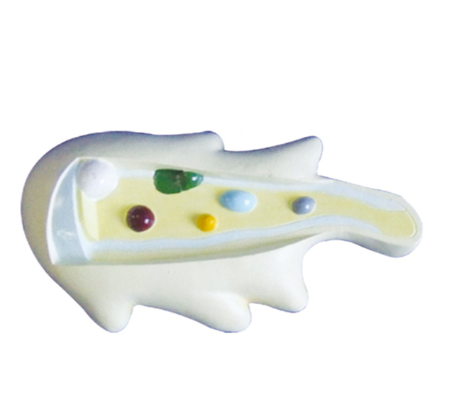
2. The groove is embedded with simulated kidney, renal pelvis, renal calyx, renal calyx, and renal papilla etc.
3. Using a clinical ultrasound diagnostic device to scan the kidneys, the outline of the kidneys, renal pelvis, kidney calices, kidney stones, etc. can be seen on the display screen.
4. The material of this model allows for translucency of ultrasound and X-rays, as well as realistic renal morphology and the location of hydronephrotic renal calices under ultrasound and X-rays.
5. According to the formal ultrasound and X-ray guided percutaneous renal puncture process, the puncture can be successfully completed, which is similar to the real surgery. Percutaneous renal puncture, guide wire insertion, channel expansion, endoscopic observation, and even lithotripsy can be performed under ultrasound guidance. It almost covers every operational step of percutaneous nephroscopy.
6. After successful puncture, simulated urine can be extracted from the needle tail.
7. The resistance felt during the expansion process of the fascial expander after inserting a guide wire is similar to that of human tissue. The degree of expansion can be determined by injecting water into the model.
8. Stones can be inserted for subsequent endoscopic observation and combined with ultrasound, pneumatic cannulation, and holmium laser energy to complete related procedures such as stone fragmentation.
Previous Page
Recommended Products
Welcome


